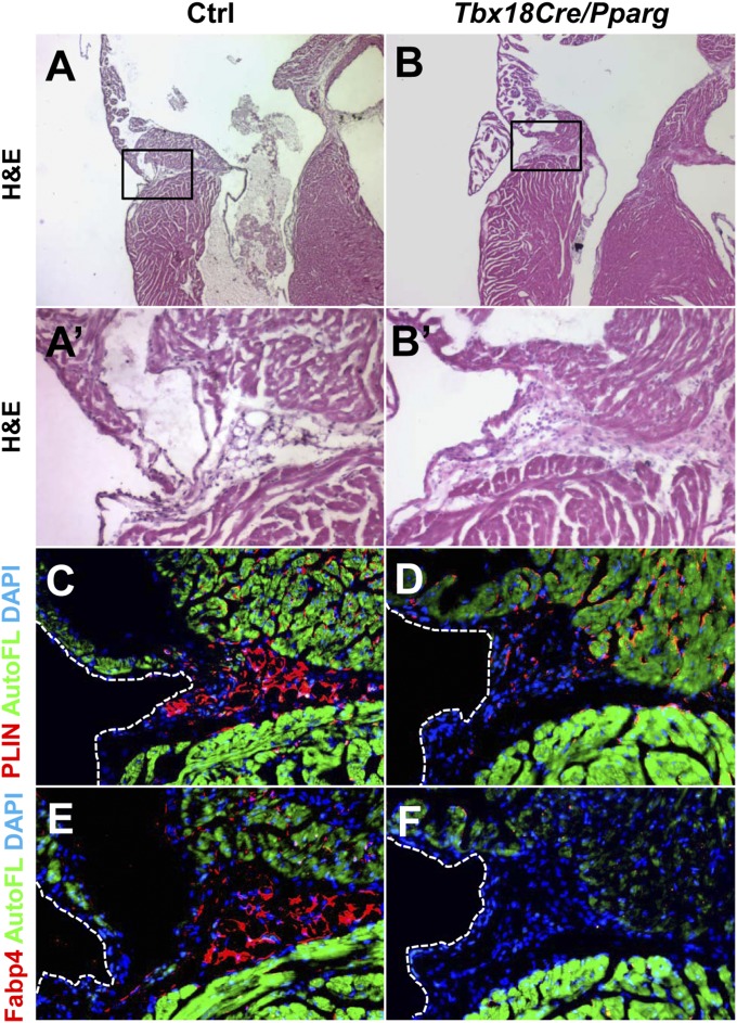
Fig. 3.
PPARγ is required for development of EAT. Sections through the AV groove of 6-wk-old mice are shown, stained either by H&E (A and B) or nearby sections stained for PLIN (C and D) or FABP4 (E and F). The locations of the high magnification views of the AV groove shown in A′, B′, and C–F are indicated by the boxed region in A and B. The scattered PLIN+ cells seen in Tbx18Cre/Pparg hearts are of unknown fate but are not adipocytes, as evidenced by their morphology and lack of staining by FABP4. Green is autofluorescence of the myocardium. Control mice (n = 4) were littermates of the Tbx18Cre/Pparg mice (n = 4).

