Fig. 4.
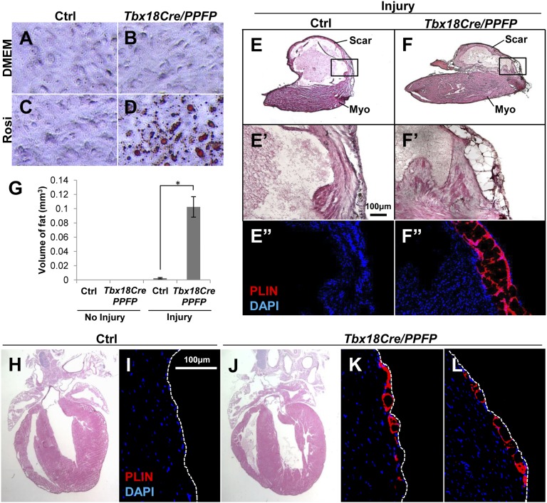
PPARγ activity and mesenchymal transformation induce adipogenesis in vivo. (A–D) In vitro induction of adipogenesis by PPFP. Primary embryonic ventricular epicardial cells were derived from Tbx18Cre/PPFP embryos and control littermates, treated with rosiglitazone, and lipid accumulation visualized by Oil Red O staining. (E and F) In vivo adult adipogenesis. Cryoinjury was performed on Tbx18Cre/PPFP adult mice and controls; a high fat diet and rosiglitazone were provided for 3 mo after surgery. In transverse sections near the apex, ventricular adipocytes were detected by PLIN immunostaining and DAPI counterstaining. Images of uninjured hearts are in Fig. S6 C and D. The boxed areas at the transition between normal myocardium (myo) and scar tissue are shown in E′, E′′, F′, and F′′; E′ and E″, and F′ and F″, are adjacent or nearby sections. (G) Quantitation of ventricular fat. The volume of ventricular fat per heart was derived from PLIN- and Oil Red O-stained serial sections. There was no fat in uninjured control (n = 4) or uninjured Tbx18Cre/PPFP (n = 5) hearts, a small amount in injured controls (n = 4), and substantially more (*P = 0.0004) in injured Tbx18Cre/PPFP (n = 5) hearts. Fat was only present in injury-adjacent areas. (H–L) Induction of embryonic adipogenesis. Pregnant females were fed a high fat diet and provided with rosiglitazone starting at E10.5 and continuing during lactation, and weaned pups were kept on the same regimen until 3 mo of age. The H&E images (H and J) show normal morphology of the treated adult hearts; immunostaining for PLIN (I, K, and L) shows the presence of adipocytes in two different locations of the ventricle in Tbx18Cre/PPFP mice but not in controls. These observations were repeated in six Tbx18Cre/PPFP mice and four control littermates (bearing only Tbx18Cre, or only PPFP, or neither allele).

