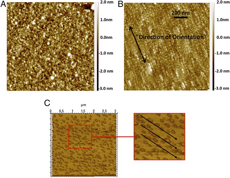
Fig. 2.
Comparison of AFM image of antibody coated surface when the field is (A) off to when the field is (B) on during IgG immobilization step. (C) AFM image of antibody coated surface when a 1,000 times diluted IgG sample was used, indicating alignment at a molecular level. The results indicate that the surface-bound antibodies were oriented in a uniform direction when the field was applied during the immobilization step.

