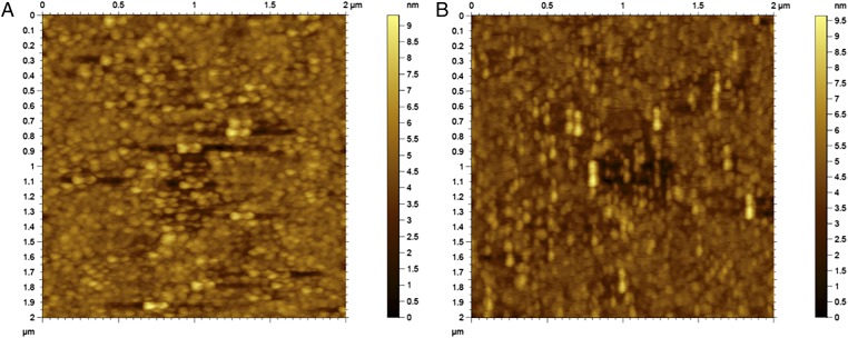
Fig. 3.
Comparison of AFM image of antibody coated surface where the AFM imaging was done at (A) 0° and (B) 90° with respect to the direction of the originally applied 8 V/cm electric field during the IgG immobilization step. This illustrates that the observed orientation of molecules is not due to AFM stamping artifact.

