Fig. 1.
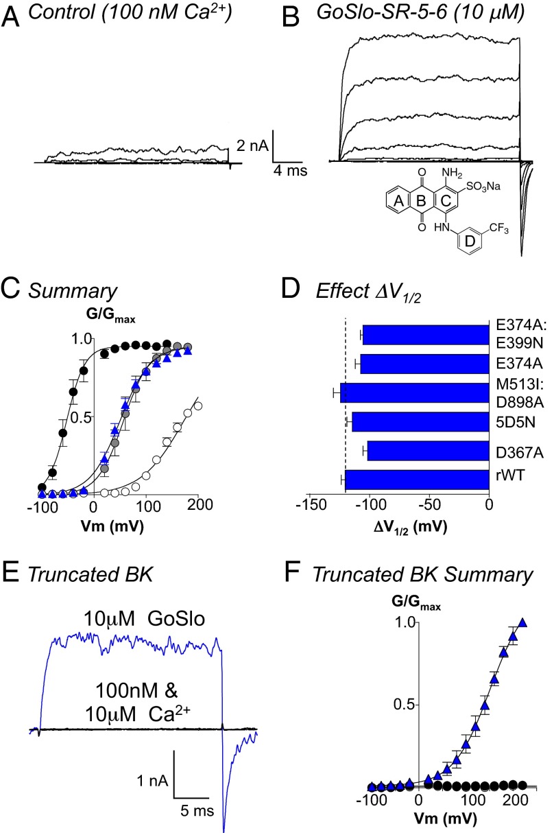
GoSlo does not need functional Ca2+ or Mg2+ sensors. A shows currents evoked from −100 to +100 mV in 20-mV steps from a holding potential of −60 mV in 100 nM Ca2+. (B) Currents evoked from the same patch following application of 10 μM GoSlo to the cytosolic face of the channel. GoSlo structure shown Inset in B. (C) Summary GV curves in 100 nM Ca2+ before (open circles) and during GoSlo (blue triangles), 1 μM Ca2+ (gray circles), and 10 μM Ca2+ (black circles), and solid lines show Boltzmann fits to the data. Data are quoted as the mean ± SEM (n = 13). (D) Mean ΔV1/2 (±SEM) of GoSlo obtained in a variety of mutants designed to reduce (D367A, n = 6; 5D5N, n = 5) or ablate (M513I:D898A, n = 6) Ca2+ or Mg2+ sensing (E374A, n = 7; E374A:E399N, n = 6). None of the mutants significantly reduced ΔV1/2. (E) The truncated BK construct [Slo1C-Kv-minT (18)] failed to evoke currents when depolarized to +100 mV and was insensitive to Ca2+ (black traces). Large currents were evoked in GoSlo (blue trace). (F) Summary GV curves for the truncated channel (n = 5) in the presence of 100 nM Ca2+ (white circles), 10 μM Ca2+ (black circles), and 10 μM GoSlo (blue triangles).

