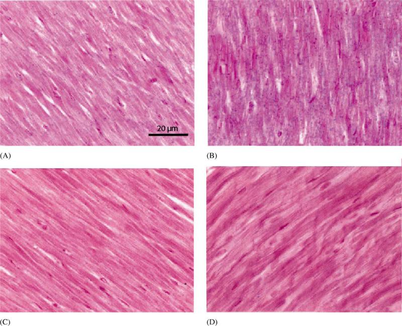
Fig. 3.
Detection of nitrotyrosine in myocardium subjected to an ischemia–reperfusion sequence after SNAP infusion. Nitrotyrosine is indicated by blue stain. (A) non-risk area; (B) risk area; (C) risk area, no primary antibody; (D) control animal (no SNAP given). Sections are counterstained with nuclear fast red. Sections are representative of four of the five animals studied. Nitrotyrosine staining was markedly greater in the risk area (ischemicre-perfused myocardium).

