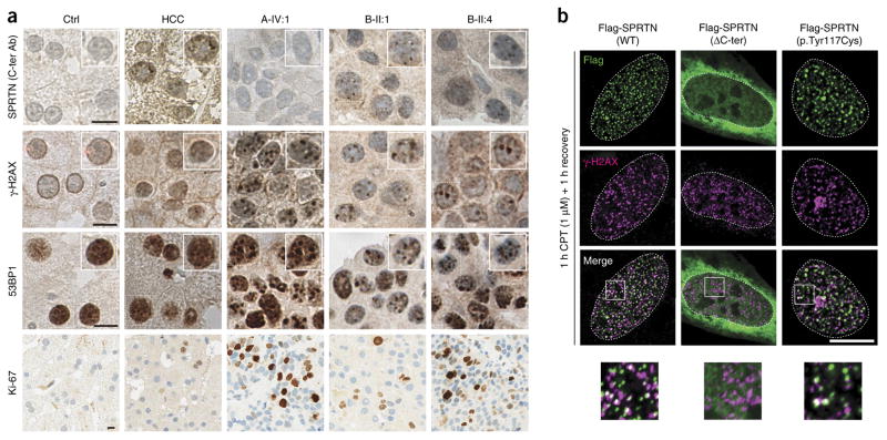
Figure 2.
Severe DNA damage in hepatocellular carcinoma biopsies and focal nuclear accumulation of SPRTN. (a) Histological and immunohistochemical analyses of human liver biopsies from a healthy control (Ctrl), a patient with idiopathic, non–viral caused HCC and the HCC of patients with SPRTN mutations (A-IV:1, B-II:1 and B-II:4). The samples were stained with antibody raised against the C-terminal part of SPRTN (C-ter Ab) or with antibodies against γ-H2AX, 53BP1 or Ki-67. The insets in the top three rows are at 1.25× magnification. (b) U2OS cells were transiently transfected with Flag-tagged WT or mutant SPRTN and challenged with 1 μM of CPT to induce replication-related DSBs and thus mimic the DNA damage observed in patients’ livers. The images at the bottom of b are 3× magnified versions of the boxed areas in the merged images above. Scale bars, 10 μm (a,b).

