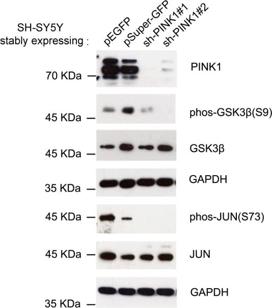
Fig. 5.
Validation of protein phosphorylation changes. (a) SH-SY5Y cells stably expressing either pSuper-GFP vector or pEGFP vector were used as control cells to compare with SH-SY5Y cells expressing pSuper-GFP containing shRNA against PINK1 at two different exonic locations. Western blot analysis of the cell homogenates using antibodies against either non-phosphorylated or phosphorylated GSK3β and c-Jun. Stable cell lines expressing different plasmids are designated.

