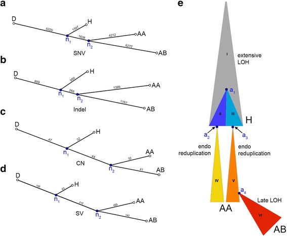
Figure 3.

Genomic lineage of tumor subpopulations. (a-d) To visualize the genetic distance between tumor subpopulations, neighbor-joining trees were constructed from distance matrices using different classes of somatic mutations. (a) Single nucleotide variant tree. (b) Indel tree. (c) Copy number aberration tree. (d) Structural variant tree. (e) Diagram of one potential evolutionary history that can explain the patterns of variant sharing shown in Figure 2a. Different clonal lineages inferred by Ploidy-Seq are indicated by distinct colors and roman numerals. Note that the relative area of each clone is not representative of the actual abundance in the analyzed tumor. Key cellular ancestors are indicated by the labels a1-a4: a1 is the last common ancestor of the genetically distinct hypodiploid clones II and III; a2 and a3 are the cells in which endoreduplication occurred to produce clone IV and clone V, which comprise the AA population; a4 is the cell giving rise to AB (clone VI), which is the most highly mutated subpopulation and appears to have undergone rapid clonal expansion.
