Fig. 4.
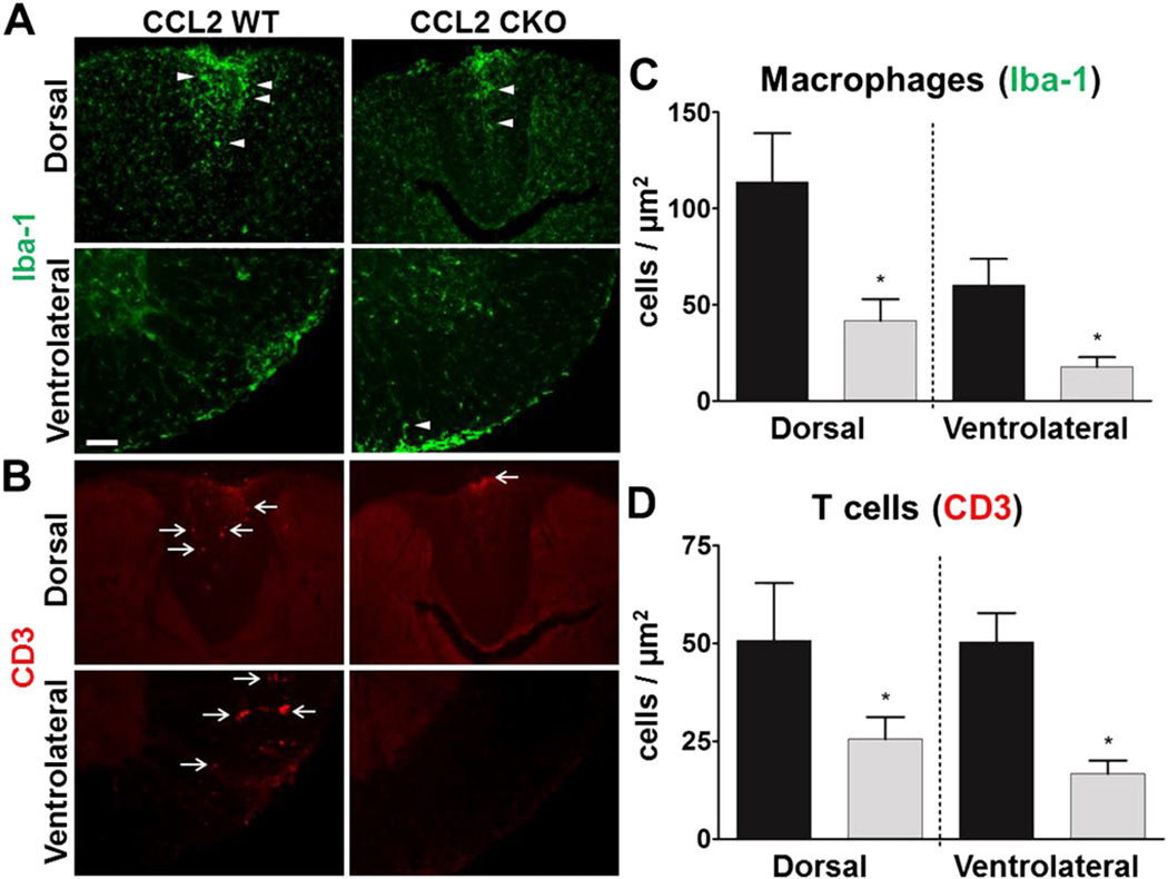
Astrocyte CCL2 CKO mice with EAE have a reduction in macrophage and T cell infiltrates in both dorsal and ventral white matter of the spinal cord during EAE. Representative immunofluorescence images of A, Iba-1 (green) globoid macrophage (arrowhead) and B, CD3 (red) T cell (arrow) infiltrates, each reduced in the dorsal (top rows) and ventralateral (bottom rows) white matter of spinal cords during EAE. Scale bar, 100 µm. Quantitative analysis show that astro-CCL2-CKO mice have a significant reduction in C, Iba-1 globoid macrophages and D, CD3 T cells in the dorsal and ventralateral white matter of the spinal cord during EAE. *p < 0.05 versus CCL2 WT (mean ± SEM).

