Figure 1.

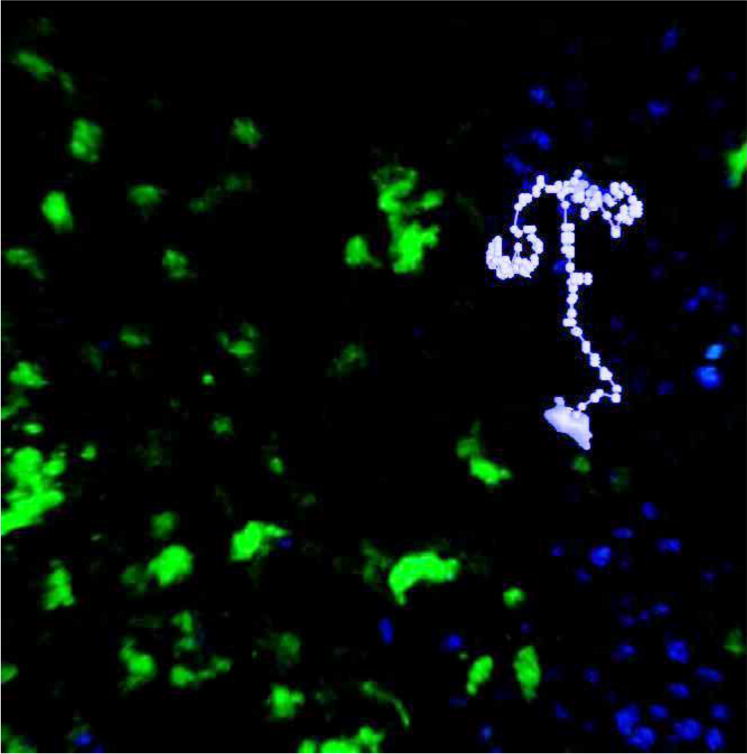
An image from a two-photon experiment overlaid with an example cell track from animal 1: Points indicate the cell centroid at 15-second imaging intervals with the entire cell shown at the beginning of the track.

An image from a two-photon experiment overlaid with an example cell track from animal 1: Points indicate the cell centroid at 15-second imaging intervals with the entire cell shown at the beginning of the track.