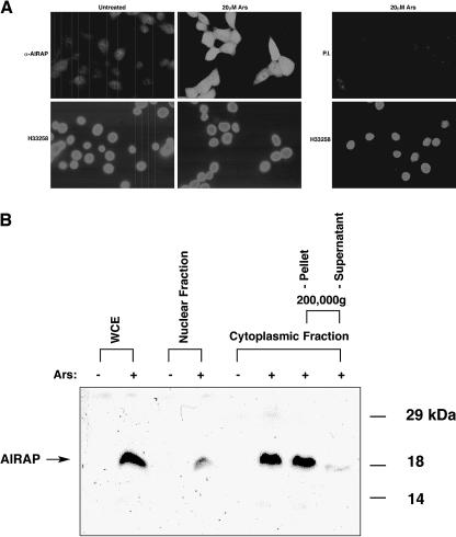
Fig 3. Detection of the endogenous arsenite-induced AIRAP protein in NIH3T3 cells. (A) Immunofluorescent localization of AIRAP in untreated cells and cells exposed to 20 μM sodium arsenite for 6 hours. Photomicrographs of fixed cells stained with rabbit anti-AIRAP serum (α-AIRAP) or preimmune serum (P.I.) and a secondary fluorescein-labeled goat anti-rabbit serum are shown. The karyophilic dye H33258 labels all the cells. (B) Immunoblot detection of AIRAP. Whole cell extracts (WCE), nuclear fraction, cytoplasmic fraction, and 200 000 × g ultracentrifugation supernatant and pellet from untreated (−) and arsenite-treated (30 μM, 6.5 hours) (+) cells were resolved by 12% SDS-PAGE and immunoblotted with the anti- AIRAP antisera. The arrow indicates the 19-kDa AIRAP protein

An official website of the United States government
Here's how you know
Official websites use .gov
A
.gov website belongs to an official
government organization in the United States.
Secure .gov websites use HTTPS
A lock (
) or https:// means you've safely
connected to the .gov website. Share sensitive
information only on official, secure websites.
