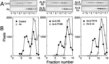
Fig 5. Western blot analysis of Hsp27 in fractions separated by centrifugation. U251 MG cells were exposed to sodium arsenite with or without protein kinase inhibitors, and extracts were fractionated by centrifugation on a sucrose density gradient as described in the caption to Figure 1. The 7.5-μL aliquots of each fraction were subjected to SDS-PAGE and subsequent Western blot analysis with antibodies against human Hsp27 and peroxidase-labeled antibodies against rabbit IgG as second antibodies as described in “Materials and Methods.” (A) Peroxidase activity on nitrocellulose sheets was visualized on X-ray film. (B) The relative density of each band in each treatment shown in (A) was estimated by the NIH Image program. Control, untreated control cells. The data are representative of 2 separate experiments

An official website of the United States government
Here's how you know
Official websites use .gov
A
.gov website belongs to an official
government organization in the United States.
Secure .gov websites use HTTPS
A lock (
) or https:// means you've safely
connected to the .gov website. Share sensitive
information only on official, secure websites.
