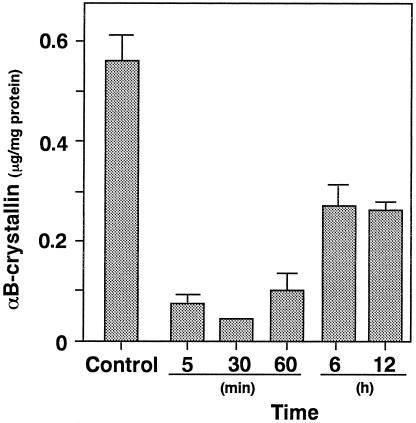
Fig 6. Levels of αB-crystallin in the vessel wall after endothelial injury. Animals were divided into a noninjured control group (n = 8) and injured group (n = 8). The injured carotid arteries were removed 5, 30, and 60 minutes and 6 and 12 hours after the initiation of injury. Each value represents the mean ± SEM

An official website of the United States government
Here's how you know
Official websites use .gov
A
.gov website belongs to an official
government organization in the United States.
Secure .gov websites use HTTPS
A lock (
) or https:// means you've safely
connected to the .gov website. Share sensitive
information only on official, secure websites.
