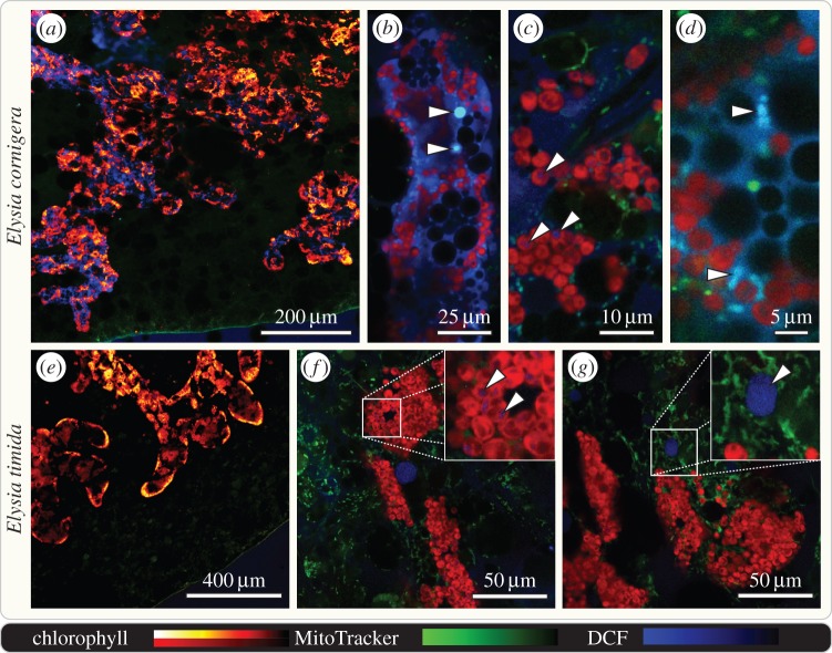
Figure 4.
Hydrogen peroxide accumulates in the digestive tubules of starving E. cornigera, but not E. timida. Confocal laser scanning micrographs of the kleptoplast-bearing digestive tubules that pervade the parapodia of 10 days starved, whole mount E. cornigera and E. timida stained with 100 µM dichlorofluorescin (DCF; blue) and MitoTracker Red (green); chlorophyll autofluorescence in red to yellow (a,e) or red only (b–d, f, g). Digestive tubules of E. cornigera (lined with autofluorescent kleptoplasts) show well-defined DCF staining within the lumen and epithelium (a–d). Details of single digestive tubules revealing readily H2O2 accumulators of equal size as kleptoplasts (b arrowheads), kleptoplasts displaying weak DCF staining (c; see arrowheads) and MitoTracker Red co-localizing with DCF staining (d). Digestive tubules of E. timida displaying no distinguishable DCF signal in epithelium or lumen (e). Details of single digestive tubules revealing accumulating weak levels of H2O2 (f, arrowheads), comparable to those seen in E. cornigera (c, arrowheads) and showing globular structures of unknown nature accumulating H2O2 (g, arrowhead).

