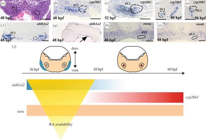
Figure 2.
Retinoic acid signalling in the tooth-forming region. (a) Transverse sections of the posterior ventral pharynx of a 48 hpf Danio rerio embryo stained with haematoxylin and eosin. The 4V1 tooth germs are marked by dashed lines. (b–e) Transverse sections of the posterior ventral pharynx of embryos post in situ hybridization embryos stained with cyp26b1 at (b) 48 hpf, (c) 52 hpf, (d) 80 hpf and (e) 96 hpf. The 4V1 tooth bud is marked by dashed lines in (b,c). The 3V1 and 4V2 tooth buds are marked by dashed lines in (d) and (e), respectively. (f) Transverse and (g) longitudinal sections of the posterior ventral pharynx of an embryo post in situ hybridization embryos stained with aldh1a2 at 48 hpf. 4V1 is marked in (f), whereas expression in the ventral posterior pharynx in the vicinity of the forming tooth bud is indicated by an arrow in (g). Note that aldh1a2 expression is excluded from the developing tooth bud in (f). (h) raraa and (i) rarab expression in transverse sections of the posterior ventral pharynx at 48 hpf. The position of the 4V1 tooth bud is indicated. Note that both raraa and rarab are expressed within the tooth bud but also in the entire posterior ventral pharynx. (j) Summary of the temporal and regional expression pattern of RA signalling actors: at 36 hpf, aldh1a2 is expressed in the ventral pharynx until 48 hpf. cyp26b1 expression starts at 50 hpf in the tooth bud mesenchyme. raraa and rarab are expressed through the entire ventral pharynx. The RA availability within the tooth-forming region is temporally sharpened by the expressions of both synthetizing (aldh1a2) and degrading (cyp26b1) enzymes. Scale bars, 25 µm.

