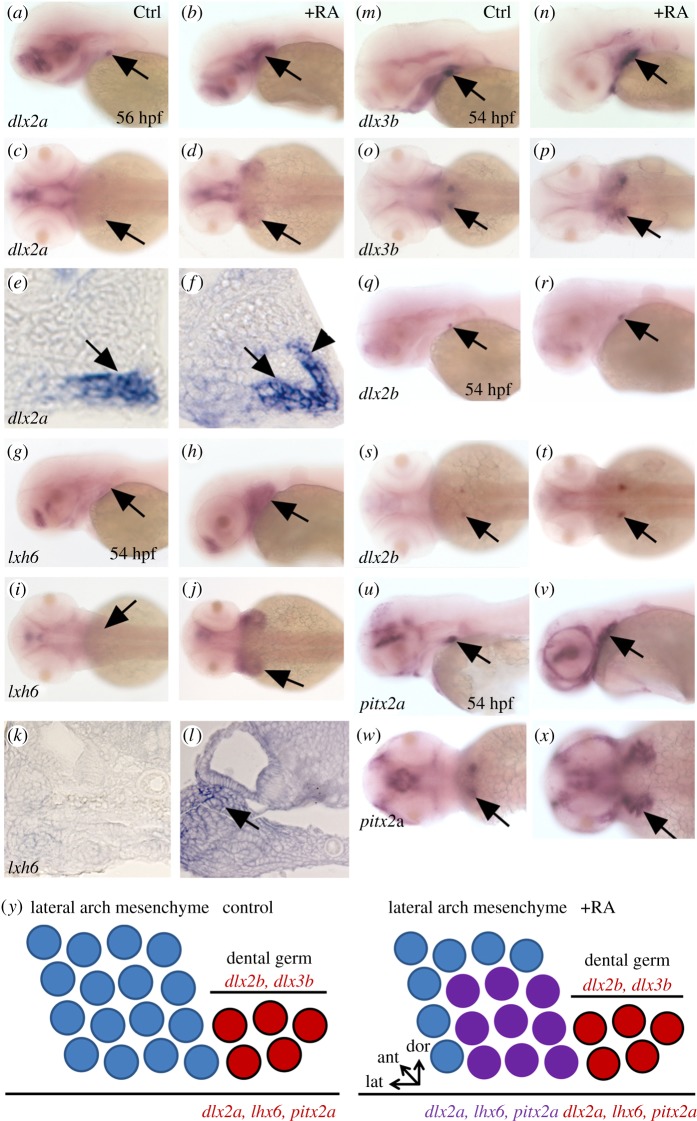
Figure 4.
Expansion of pharyngeal mesenchyme markers in RA treated embryos. (a–f) expression of dlx2a in the tooth bud of 4V1 and lateral arch mesenchyme marked by an arrow in (a,c,e) control and (b,d,f) RA-treated embryos. Note the lateral, anterior and dorsal expansion of dlx2a expression in RA-treated embryos (arrowhead in (f)). (g–l) Expression of lhx6 in the lateral pharyngeal mesenchyme marked by an arrow in (g,i,k) control and (h,j,l) RA-treated embryos. As for dlx2a, note the lateral, anterior and dorsal expansion of lhx6 expression in RA-treated embryos (arrow in transverse section in (l)). (m–t) Expression of dlx2b and dlx3b in the dental epithelium marked by an arrow in (m,o,q,s) control and (n,p,r,t) RA-treated embryos. RA exposure has only a minor effect on the expression of these dental epithelium markers. (u–x) Expression of pitx2a in the pharyngeal and dental epithelium marked by an arrow in (u,w) control and (v,x) RA-treated embryos. Note the lateral, anterior and dorsal expansion of pitx2a expression in RA treated embryos. (y) Diagram of a longitudinal section of the posterior ventral pharyngeal epithelium and mesenchyme of the 4V1 tooth germ during early morphogenesis, dorsal up, anterior to the left of a control embryo (top) and RA-treated embryo (bottom). In red, dlx2b and dlx3b are restricted to the folding dental epithelium, whereas dlx2a is expressed in both the developing tooth (red) germ and the lateral pharyngeal mesenchyme (purple); lhx6 is expressed in both the dental mesenchyme (red) and in lateral arch mesenchyme (purple); pitx2a is expressed in the dental epithelium (red) and the pharyngeal epithelium (purple). Under RA exposure, expression of dlx2a, lhx6 and pitx2a is expanded in three directions (laterally, dorsally and anteriorly), whereas expression of dlx2b and dlx3b remains confined in the dental epithelium, but their expression domains are slightly enlarged compared with control.

