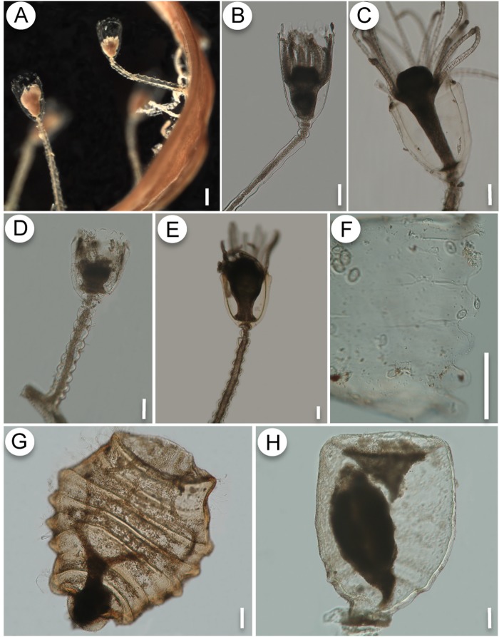
Fig 6. A-C, G: Orthopyxis sargassicola.
A: general view of the colony on Sargassum sp.; B-C: detail of the trophosome, showing variation in perisarc thickness of hydrotheca; G: gonotheca. D-F, H: Orthopyxis crenata. D-E: detail of the trophosome; F- detail of the hydrothecal cusps; H- gonotheca. Scales: A—200 μm; B-H—100 μm.

