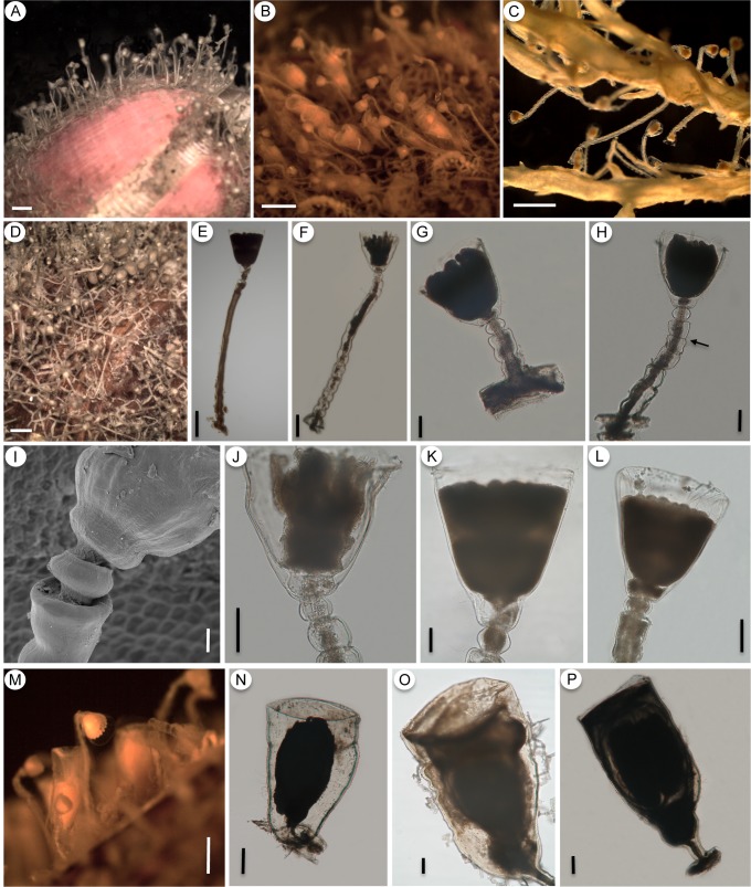
Fig 9. Orthopyxis mianzani sp. nov.
A-C: general view of the colony (A-MZUSP 2575; B-MZUSP 2580; C-MZUSP 2559); D: detail of the hydrorhiza (USNM 1259970); E-H: details of the trophosome, showing variation in pedicels from smooth (E) to sinuous (F), variation in the length of the pedicels (compare E, F and G) and constrictions of the perisarc (H, arrow) (E-MZUSP 2576; F-MZUSP 2572; G-MZUSP 2570; H-MZUSP 2574); I: detail of subhydrothecal spherule (USNM 1259970); J-L: detail of hydrothecae (J-MZUSP 2572; K-MZUSP 2576; L-MZUSP 2579); M: general view of the gonotheca on natural substrate (MZUSP 2580); N-P: detail of female gonothecae (N-MZUSP 2572; O-USNM 1259970; P-MZUSP 2580). Scales: A-D—1 mm; E—300 μm; F, N, P—200 μm; G, H, J-L, O—100 μm; I—20 μm; M—500 μm.

