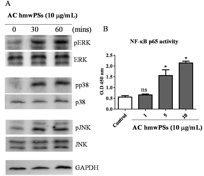
Fig 5. Activation of MAPKs and NF-κB by AC hmwPSs in DCs.
DCs were collected and treated with AC hmwPSs (10 μg/mL). Lysis buffer was added at the indicated time points to obtain whole cell lysates or nuclear extracts. (A) The activation of MAPKs was assayed by western blot. Whole cell lysates were incubated with antibodies against phosphorylated protein of ERK, JNK, and p38 MAPK, and then detected with specific 2nd antibodies and ECL system. The loading controls are shown and represent the use of antibodies against the total protein of these MAPKs. The data are representative of three independent experiments. (B) The activation of NF-κB was determined by p65 binding assay. The NF-κB binding activity in nuclear extract was measured using TransAM NF-κB p65 ELISA kit and shown as value read at OD450. The data are the mean + SD of three samples. NS p>0.05 (Mann–Whitney U test) are comparisons between LPS- or AC hmwPS-treated and PBS-treated DCs. All of the results are representative of three independent experiments.

