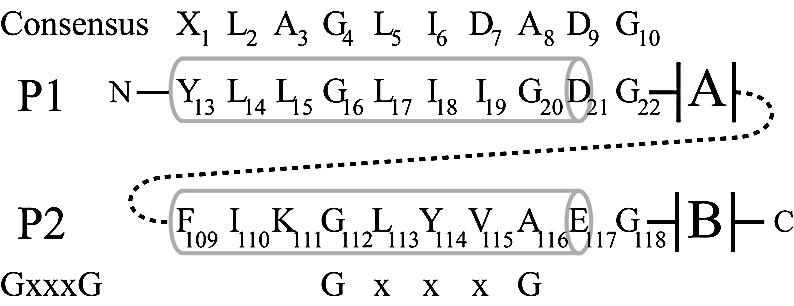
Figure 1.

Schematic alignment of the LAGLIDADG residues of I-DmoI. Pertinent residues in P1 and P2 are identified below the consensus LAGLIDADG sequence. The alignment of the GxxxG motif is shown on the bottom. Numbers in the consensus sequences represent generic positions within the LAGLIDADG motif. Numbers in P1/P2 represent positions of residues in I-DmoI. Gray barrels indicate helical structure. A and B designate the two α/β domains that form the bulk of the protein. Dashed line represents the short (six to eight amino acids) linker between the subdomains A and B in I-DmoI.
