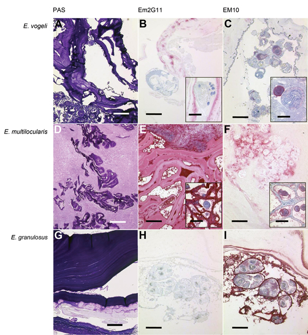
Figure.

Periodic acid–Schiff (PAS) staining and immunohistochemical analysis of samples from Echinococcus vogeli lesions, with monoclonal antibodies against Em2 and EM10 of E. vogeli (A, B, C), E. multilocularis (D, E, F), and E. granulosus (G, H, I) lesions. Staining was performed on archived tissue from human patients with alveolar and cystic echinococcosis for comparison, and from the patient with E. vogeli infection who immigrated to the Netherlands from Suriname (E. vogeli infection in 2009). B and C insets) Protoscolex, with rostellar hooks clearly visible in inset B. E and F insets) Tissue from infected rodents (laboratory-infected Meriones unguiculatus gerbils) because E. multilocularis lesions in humans only rarely contain protoscolices. A, D, G) PAS-stained sections of the respective echinococcal lesions. Scale bars indicate 500 μm. B, E, H) Lesions with E. vogeli, E. multilocularis, and E. granulosus infection, respectively, stained with the monoclonal Em2G11 antibody against Em2 (for staining details see [7]). E. multilocularis lesions show intense staining, E. granulosus lesions show no staining, and E. vogeli lesions show patchy stains. Scale bars indicate 200 μm; scale bars of the insets indicate 50 μm. C, F, I) Respective lesions stained with antibodies against EM10 (dilution of the primary antibody 1:50; further steps as in Barth et al. [7]). Germinal layer and protoscolices of E. multilocularis and E. granulosus larvae are stained, but the protoscolices of the E. vogeli metacestode are only partly stained. Scale bars indicate 200 μm; scale bars of the insets indicate 50 μm.
