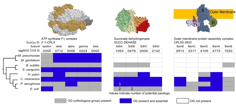
Fig 4. Examples of protein complex conservation across bacteria.
Complexes are identified using a common name and an EcoCyc ID. Each complex subunit has been assigned a COG ID. Grey areas indicate OG presence, white areas indicate OG absence, and blue areas indicate essentiality in a species-specific screen (see Materials and Methods for references). Values within these spaces indicate the presence of potential paralogs in the corresponding species; proteins without specified values have no paralogs. Complex structures are available in PDB: ATP synthase F1, 3OAA; succinate dehydrogenase, 1NEN; Outer membrane protein assembly complex, (2KM7, 3TGO, 3TGO, 4K3C, 2YH3). Species are arranged by their taxonomy (see Materials and Methods for details) with E. coli and Mycoplasma serving as the most distant species.

