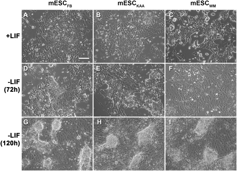
Fig 3. Morphology of mESC.
(A-C) Undifferentiated stem cells. (D-F) 72h after LIF removal. (G-I) 120h after LIF removal. Embryoid bodies are clearly visible at this stage. All photographs were taken under a 10X objective and the scale bar represents 20μm. No differences in morphology between conditions were observed at any time points.

