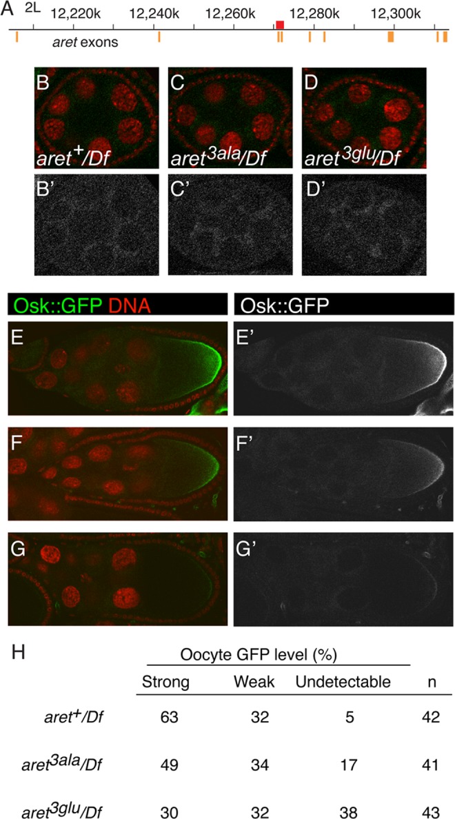
Fig 7. Translational repression and activation in aret mutants.

A. Schematic diagram of the aret locus. Numbering is according to the Drosophila genome sequence, R5.48. Orange bars depict exons (the widths of the bars are not to scale), and the red rectangle is the 2.1kb targeted region, which when translated, includes the amino-terminal domain of female Bru.B-D, B’-D’. Stage 6 egg chambers expressing the osk1–534::GFP-AB reporter in different aret mutant backgrounds, as labeled. Df is Df(2L)BSC407. For B-D GFP is in green and nuclei in red. Panels B′-D′ show just the GFP channel. E-G, E’-G’. Examples of phenotypic categories for panel H. For E-G GFP is in green and nuclei in red. Panels E′-G′ show just the GFP channel. All samples were fixed in parallel and imaged together under the same settings. Expression of the UAS transgene was driven by the matα4-GAL-VP16 driver. H. Intensity of the posterior zone of GFP from UAS-osk1–534::GFP-AB in aret mutants. Examples of strong, weak and undetectable are shown in E/E′, F/F′ and G/G′, respectively.
