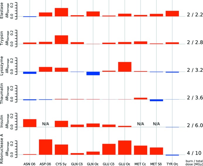
Figure 6.
Change in B Damage between low- and high-dose datasets for different residue atoms. The change in the median B Damage for ten groups of atoms of the six protein structures before and after an X-ray burn is shown. Insulin does not have ASP or MET residues. Different proteins show different B Damage patterns. B Damage tends to increase for most atoms shown, including ASP Oδ, CYS Sγ, GLU Cδ and O∊, MET C∊ and Sδ. This indicates that these atoms damage faster than others within the same protein, and correlates well with the known specific damage decay pattern of amino acid residues.

