Figure 7.

B
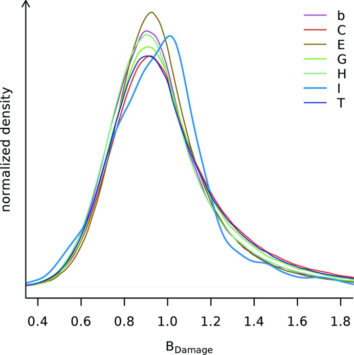
Damage distribution for different protein sections as identified by STRIDE secondary structure annotation. The seven secondary structure labels are: alpha helix (H), 3–10 helix (G), π-helix (I), extended conformation (E), isolated bridge (b), turn (T) and coil (C, none of the above) (Frishman & Argos, 1995 ▶). 33.2% of all residues within the PDB subset were marked alpha helix, 23.4% extended conformation, 19.9% turn, 17.9% coil, 4.3% 3–10 helix, 1.2% isolated bridge, and 0.016% π-helix. Applying Levene’s test with median centres (Brown & Forsythe, 1974 ▶) for equal variance on a sample of 1500 atoms from each of the seven secondary structure labels, different variances are confirmed (p <  ). All of the distributions are smoothed using a kernel density estimator (R Development Core Team, 2011 ▶).
