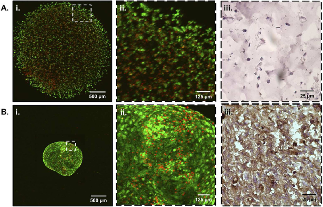
Figure 3.
LIVE/DEAD confocal microscopy of C3H10T1/2s encapsulated in PEG hydrogels (Ai) and traditional pellet cultures (Bi) both exhibit > 90% cell viability (live cells; calcein AM (green), dead cells; ethidium homodimer (red)). Cell-cell contacts are reduced in PEG hydrogel encapsulations (Aii) as compared to pellet cultures (Bii). Representative images of frozen cell-laden hydrogels (Aiii) and pellets (Biii) were immunohistochemically stained for N-Cadherin (brown) to further illustrate decreased cell-cell contact in hydrogel encapsulated MSCs.

