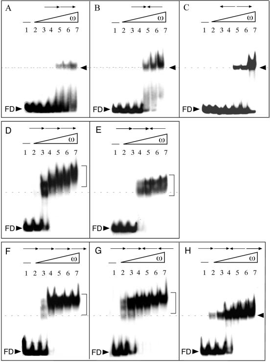
Figure 2.
Interaction of ω2 protein with DNA containing two to four heptads in different orientations. The 59 [(A–C) two heptads], 66 [(D and E) three heptads] and 73 bp [(F–H) four heptads] [α-32P]HindIII–KpnI DNA (2 nM) and 1 µg of poly[d(I-C)], as non-specific competitor DNA, were incubated with increasing concentrations of ω2 in buffer B for 15 min at 37°C. The formed ω2–DNA complexes were analyzed by EMSA. The arrows at the top of the gels indicate number and orientation of the 7 bp repeats. An arrowhead or a bracket to the right of the bands indicates the type(s) of ω2–DNA complexes. A broken line indicates the position of the fastest moving complexes, detected at low protein concentration. Except in (C), the ω2 protein concentrations were 4, 8, 16, 32, 64 and 128 nM (lanes 2–7, respectively). In (C) the ω2 concentrations were 8, 16, 32, 64, 128 and 256 nM. The symbol – in lane 1 indicates the absence of protein, FD labels the band due to free DNA.

