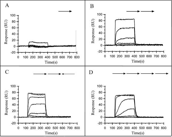
Figure 4.
SPR studies of the interaction of ω? protein with its target sites. DNA segments containing one (A), two (B), three (C) and four (D) heptads with total lengths of 20, 27, 34 and 41 bp, respectively, have been used. The nucleotide sequences are presented in Materials and Methods and number and orientation of the heptads are indicated by arrows. The sensograms were obtained at different ω2 concentrations (1.6, 3.2, 6.4, 12.5, 25, 50, 100 and 200 nM). Several curves overlap at low ω7 concentrations [three and two curves in (C) and (D), four curves in (B), and seven curves in (A)].

