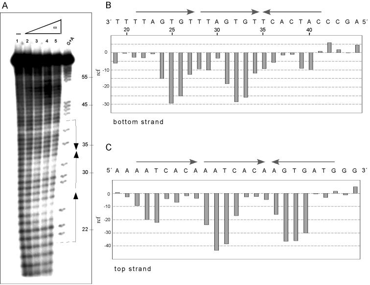
Figure 7.
Hydroxyl radical footprinting of ω2 protein bound to DNA with three heptads. The 66 bp [α-32P]HindIII–KpnI [bottom strand (A and B)] or the 71 bp [α-32P]EcoRI–SphI [top strand (C)] (2 nM) with three DNA heptads in —→2←— orientation, in the presence of 1 µg of poly[d(I-C)] as non-specific competitor DNA, was incubated and hydroxyl radical footprinted at ω2 concentrations as described in Figure 6. The nucleotide sequence of —→2←— DNA and histograms of hydroxyl radical footprinting of bottom (B) and top (C) strands are shown. The relative cleavage frequency is shown as described in Figure 6.

