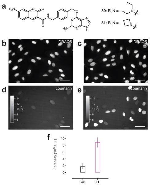
Figure 3. Utility of azetidinyl coumarins in cellular imaging.
(a) Chemical structures of commercial coumarin SnapTag ligand 30 and azetidine-containing ligand 31. (b, d) Wide-field fluorescence microscopy images of live HeLa cells expressing SnapTag–H2B and labeled with DRAQ5 and commercial SnapTag ligand 30. (b) Fluorescence of DRAQ5 nuclear staining. (d) Fluorescence of coumarin 30-labeled SnapTag–H2B. (c, e) Wide-field fluorescence microscopy image of live HeLa cells expressing SnapTag–H2B and labeled with DRAQ5 and novel azetidinyl-coumarin SnapTag ligand 31. (c) Fluorescence of DRAQ5 nuclear staining. (e) Fluorescence of coumarin 31-labeled SnapTag–H2B. Scale bars for all images: 50 μm. (f) Quantification of the average nuclear fluorescence above background coumarin label in cells when labeled with ligand 30 (black) or 31 (magenta; error bars show s.e.m.).

