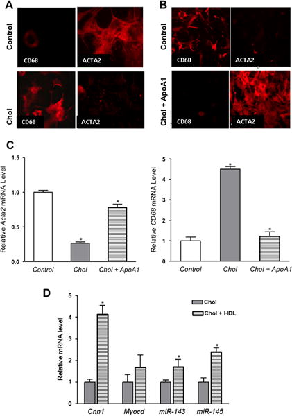
Figure 6. Cholesterol efflux promoted by apoA1 or HDL reverses phenotypic changes induced by cholesterol-loading of VSMC.

(A) Mouse VSMC were treated and immunostained as in Fig. 1. Images shown are (40×). As expected, the protein level of ACTA2 decreased, while that of CD68 increased. (B) Next, cholesterol-loaded VSMC were incubated with apoA1 for 72 h, which restored the typical VSMC appearance and ACTA2 expression, with loss of CD68 expression. (C) Consistent with immunostaining, cholesterol-loaded cells had reduced and increased, respectively, Acta2 and Cd68 mRNA levels. (D) Cholesterol-loaded VSMC were treated as in panel (C), but HDL was used instead of apoA1. By qRT-PCR analysis, there were recoveries of the expression of the VSMC marker Cnn1 as well as of the regulatory factors Myocd and miR-143/145. *p < 0.5 vs. control). Data shown are mean ± SD of triplicates of qRT-PCR reactions and are representative of two independent experiments.
