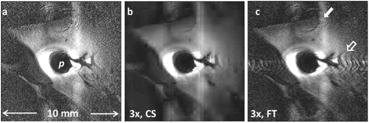
Fig.4.

(a) Regular Cartesian in vivo MRI endoscopy of a rabbit aorta acquired with a transmit/receive loop coil (8). (b) Three-fold under-sampling using CS reconstruction (SSIM vs. 4a =0.53). (c) The under-sampled k-space from (b), when conventionally reconstructed, shows strong artefacts (hollow arrow) near the probe. The vertical bright band (arrow) is likely due to motion which cannot be corrected in this Cartesian acquisition (SSIM vs. 4a =0.69).
