
Fig.9.
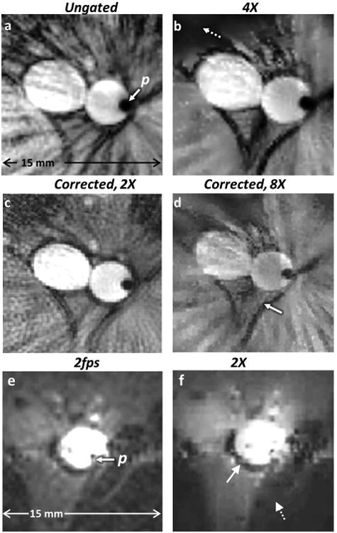
(a) Ungated radial IVMRI (zoomed anatomy of Fig. 8b) with conventional scanner reconstruction using even and odd spokes shows streaking near the aorta and surrounding tissue. (b) Tissue morphology is retained in a 4-fold under-sampled CS reconstruction of part (a) albeit with some loss of signal in regions of low SNR (dashed arrow; SVratio vs. 9a =0.9). (c) Motion-correction applied to half the data (using only odd spokes) in part (a) with conventional NUFFT reconstruction reduces streaking (SVratio vs. 9a =0.95). (d) Eight-fold under-sampling of part (a) using only odd spokes, retains morphology of the aorta and surrounding tissue (solid arrow), although the intensity of the blood signal is reduced (SVratio vs. 9c =1.1). (e) IVMR images from a rabbit aorta in vivo, acquired at 2fps without any cardiac gating. (f) Two-fold under-sampled CS reconstruction without any motion correction retains overall morphology (aorta, solid arrow), but some detail is lost in low SNR regions (dashed arrow). The r-1 intensity filter has not been applied to (e) or (f) (SSIM of 9f vs. 9e =0.82).