Figure 1.
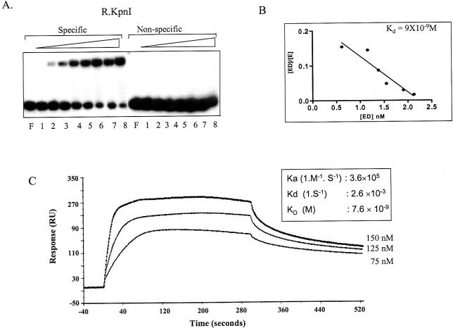
KpnI REase DNA binding. (A) Electrophoretic mobility shift assay. Labeled specific and non-specific 38mer oligonucleotides were used for the binding assay. KpnI REase: 10 nM of labeled oligonuclotides were incubated with 0.05, 0.25, 1.25, 6.5, 32, 64, 125 and 250 nM (lanes 1–8) of KpnI REase. Lane F indicates free DNA. (B) Scatchard analysis of KpnI REase DNA binding data from (A). (C) SPR analysis of the interaction of KpnI REase at its recognition sequence. The different concentrations of KpnI REase ranging from 75 to 150 nM were injected for 300 s over the SA chip immobilized with biotinylated oligonucleotide at a flow rate of 10 µl/min followed by a dissociation phase (300 s). The kinetic parameters are given in the table (inset).

