Figure 3.
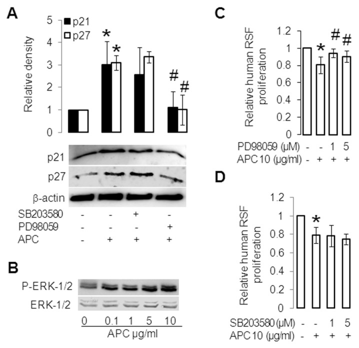
APC inhibits proliferation via ERK in RSFs. (A) RSFs were pretreated for 1 h with SB203580 (10 μmol/L) or PD98059 (10 μmol/L) and then treated without (control) or with APC (10 μg/mL) for 24 h. p21 and p27 were assessed by Western blotting. β-Actin was used as a loading control. The band intensity of the proteins was normalized with β-actin, and controls were defined as 1. Values are shown as mean ± SD; n = 3; *p < 0.05 versus control; #p < 0.05 versus APC treated RSFs, with the use of paired t test. (B) RSFs were treated without or with APC for 30 min; ERK1/2 and phosphorylation (P) of ERK1/2 were assessed by Western blotting. (C, D) RSFs were pretreated for 1 h without or with PD98059 (1, 5 μmol/L) (C) and SB203580 (1, 5 μmol/L) (D) and then treated with or without APC (10 μg/mL) for 24 h. Proliferation was measured by crystal violet assay. Controls were defined as 1. Values shown are mean ± SD; n = 4; *p < 0.05 versus control; #p < 0.05 versus APC-treated RSFs, with the use of a t test.

