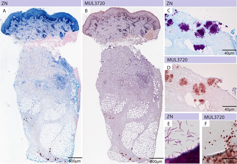
Fig 5. Immunohistochemical analysis of M. ulcerans using anti-MUL_3720 antibodies.
Histological sections were either stained with carbolfuchsin (counterstain methylenblue) (A, C, E) or with specific monoclonal antibodies (JD3.2) against MUL_3720 (B, D, F). (A) A punch biopsy of a BU patient showing the typical histopathological hallmarks of an active lesion and harbouring large clusters of AFBs inside the subcutaneous tissue (purple clumps) is depicted. (B) Staining of the same tissue specimen with anti-MUL_3720 antibodies. Serial sections were stained by ZN (C) and anti-MUL_3720 antibodies (D). While the whole bacterium is stained by ZN (E) single dots are detected by using anti-MUL_3720 antibodies (F).

