
Figure 1.
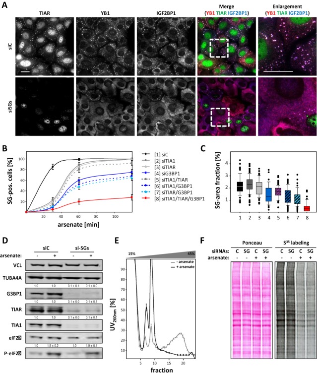
The concomitant depletion of TIA proteins and G3BP1 impairs SG formation. (A) U2OS cells transfected with indicated siRNAs (siC, control; siSG, siTIA1, siTIAR and siG3BP1) for 72 h were stressed by arsenate (2.5 μM) for 2 h before immunostaining of indicated proteins. Enlargements of the boxed regions in the merged images are shown in the right panel. Bars, 25 μm. (B and C) The average number of SG containing cells (B) and the SG area fraction (C) was analyzed by immunostaining for IGF2BP1 and YB1 in U2OS cells transfected with indicated siRNAs. Both parameters were determined by an automated particle detection tool, adapted from (18), after indicated times of arsenate stress. Error bars indicate SD determined by analyzing at least 100 cells per condition in three independent experiments. (D) The phosphorylation of eIF2α in non-stressed (−) or arsenate (+, as in A) stressed U2OS cells transfected with control (siC) or siSG (as in A) siRNAs was determined by western blotting with indicated antibodies. VCL and TUB4A4 served as loading controls to determine knockdown efficiencies as indicated by numbers above each panel. Standard deviation was determined from three independent experiments. (E) The association of bulk (m)RNA with polysomes was monitored by linear (15–45% w/v) sucrose gradient centrifugation in stressed (+, arsenate) versus non-stressed (−, arsenate) U2OS cells transfected with control (siC) or siSG siRNAs, as in (A). The distribution of RNA was monitored by UV spectroscopy and is shown as the average absorbance determined for individual fractions isolated from siC- and siSG-transfected samples. Error bars indicate SD determined in three independent studies for siC- and siSG-transfected cells. (F) Protein synthesis in arsenate-stressed (+) versus non-stressed (−) U2OS cells transfected as in (A) was analyzed by metabolic labeling using S35-methionine. The fraction of newly synthesized proteins was determined by western blotting using autoradiography (right panel). Equal loading was controlled by Ponceau staining (left panel).