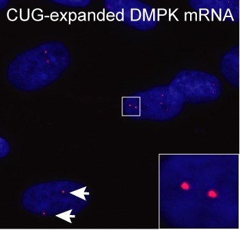
Figure 2.

CUG-foci visualized by RNA-fluorescence in-situ hybridization. CUG-expanded DMPK mRNA accumulates in distinct foci in DM1 patient cells (red foci indicated at the tip of white arrows and enlarged in inset). Blue color indicates nucleus (DAPI staining).
