
Figure 2.
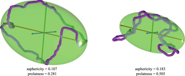
Examples of simulated momentary configurations of non-supercoiled (left) and supercoiled DNA molecules (right) together with their characteristic ellipsoids of inertia. The three principal axes of rotation a, b and c, where a ≥ b ≥ c, are shown as red, green and blue, respectively. The values of their asphericity and prolateness are indicated. The shown configurations have typical shapes obtained in simulations of diluted non-supercoiled and supercoiled (σ ≈ −0.05) DNA molecules, respectively. Their aspericity and prolatness values are both close to the corresponding average values for this type of simulated DNA molecules (see Figures 3a and c, and 4 a and c). Notice that the supercoiled DNA molecule is more aspherical and more prolate than the non-supercoiled molecule.