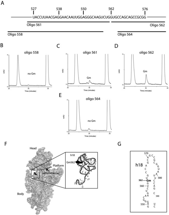
Figure 2.

Mapping of precise location of new Gm residue in the 18S rRNA. To identify precise location of the Gm residue within fragment covering nucleotides from 527 to 576 of 18S rRNA, shown in panel (A), four overlapping fragments using oligo-558, oligo-561, oligo-562 and oligo-564, each 50 nucleotides in size were isolated using mung bean nuclease assay. Nucleosides composition of these fragments was next analyzed using RP-HPLC. RP-HPLC chromatogram of oligo-558 (B), oligo-561(C), oligo-562 (D) and oligo-564 (E). The compositions of these fragments suggested G562 to contain 2′-O-ribose methylation in the 18S rRNA. Insignificant amount of Gm residue was also observed in 18S rRNA fragment isolated using oligo-561, which most likely is derived from neighboring Gm562. (F) 3D cartoon of the yeast 18S rRNA highlighting the location of Gm562 (black spheres) with a zoomed in view of helix 18. PDB file 3U5B was used for the representation of 18S ribosomal RNA. The cartoon was made by PyMol software (PyMOL Molecular Graphics System, Version 1.2r3pre, Schrödinger, LLC.). (G) 2D sequence map of the helix 18 of the yeast 18S rRNA.
