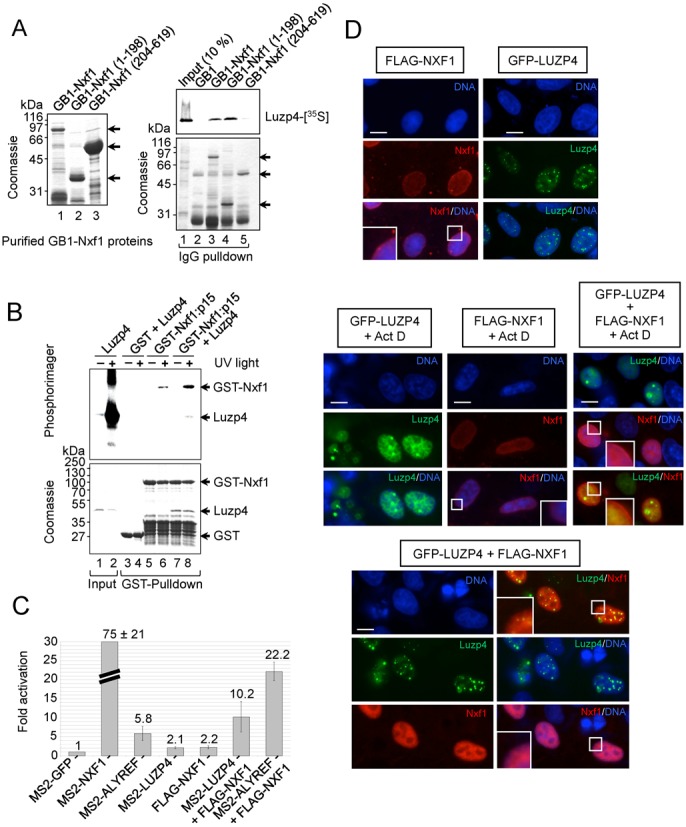
Figure 3.

Luzp4 is an mRNA export adaptor. (A) The purified GB1-tagged Nxf1 proteins used in pulldown assays are shown (left panel). IgG-Sepharose pulldown assays using 35S-Luzp4 and the indicated GB1-tagged Nxf1 proteins (right panels). The upper panel is the phosphoimage of the same gel which is Coomassie stained in the lower panel. (B) Nxf1 remodelling assays. Luzp4 pre-incubated with a 32P-labelled RNA oligonucleotide was incubated with immobilized GST or GST-Nxf1:p15. The indicated complexes were then purified by GST-pulldown, eluted, UV-crosslinked and analysed by sodium dodecyl sulphate-polyacrylamide gel electrophoresis. The upper panel is the phosphoimage of the same gel shown Coomassie stained in the lower panel. Input was 1%. (C) Tethered mRNA export assays (also see Supplementary Figure S4A). The indicated MS2 fusion protein expression vectors were co-transfected with the MS2 reporter pLUCSALRRE6MS2, normally retained in the nucleus, together with a β-galactosidase expression vector. Luciferase activities were measured from the MS2 reporter and normalized for transfection efficiency with the β-galactosidase activities. The graph shows fold activation relative to the levels seen with MS2-GFP and values represent averages from experiments carried out in triplicate on five separate occasions. (D) Immunofluorescence images of HeLa cells transfected with the indicated expression vectors. FLAG-Nxf1 was detected using FLAG antibody. When indicated, cells were treated with 5 μg/ml of actinomycin D for 3 h. Certain regions of images indicated by a white box are shown at higher magnification in the bottom left of a panel to observe the presence or reduction of FLAG-Nxf1 at the nuclear rim.
