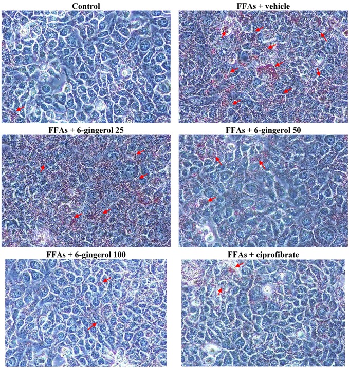
Figure 1.
Representative images of FFAs mixture-treated HepG2 cells stained with Oil Red O after different treatments. HepG2 cells were exposed to a FFAs mixture (0.33 mmol/L palmitate and 0.66 mmol/L oleate) for 24 h, and were then incubated for another 24 h with 6-gingerol at concentrations of 25 μmol/L (6-gingerol 25), 50 μmol/L (6-gingerol 50), or 100 μmol/L (6-gingerol 100), or with 100 μmol/L of ciprofibrate (ciprofibrate). The vehicle (distilled water) used to prepare the test medication solutions was given at the same volume. Cells supplemented with medium without any treatment served as control. Cells were observed by light microscopy at a magnification of 400×. Arrows indicate fat droplets.

