
Figure 4.
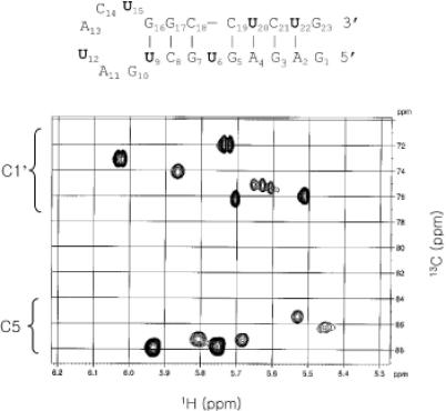
A section of the constant-time 1H–13C HSQC spectrum of the 13C,15N-UTP-specific labeled RNA recorded at 30°C. The concentration of RNA is 0.8 mM. The sample is in 99.96% D2O solvent and 10 mM phosphate buffer (pH 6.8). The 1H dimension is on the horizontal axis, and the 13C dimension is on the vertical axis. The C1′s of the uracils resonate at 72–76 ppm, and the C5s at 85–88 ppm. The 13C,15N -UTP-specific labeled U residues in the 23 nt RNA are indicated in boldface type.