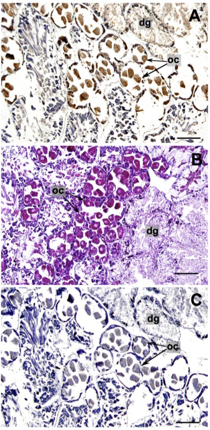
Figure 2.
Pleurobranchaea maculata gonad/digestive tissue sectioned at 10 μm. (A) Tetrodotoxin (TTX)-specific monoclonal antibody (mAB) immunohistological staining (TTX identified by the brown color deposits); (B) Hematoxylin and Eosin staining; and (C) mAB negative control. dg = digestive gland, oc = oocyte. Black bars = 200 μm.

