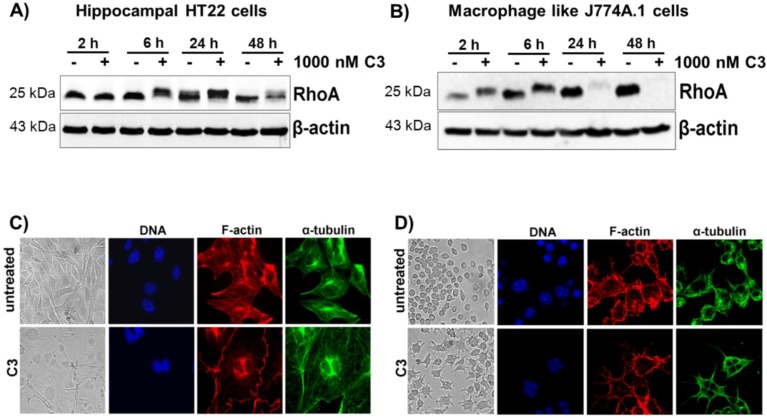
Figure 1.
Cellular susceptibility of HT22 and J774A.1 cells to C3 hippocampal HT22. HT22 cells (A) and J774A.1 macrophages (B) were treated with 1 µM of C3 for the indicated time points at 37 °C. Cell lysates were submitted to western blot analysis probing RhoA and β-actin. One representative western blot experiment is shown (n = 3). C3-induced morphological changes of HT22 cells (C) and J774A.1 cells (D) were studied by phase-contrast microscopy and confocal laser scan microscopy after 48 h of incubation. For the immunofluorescence microscopy, the cells were fixed, permeabilized and stained for α-tubulin, F-actin and nuclei. Untreated cells served as the control.

