Figure 4.

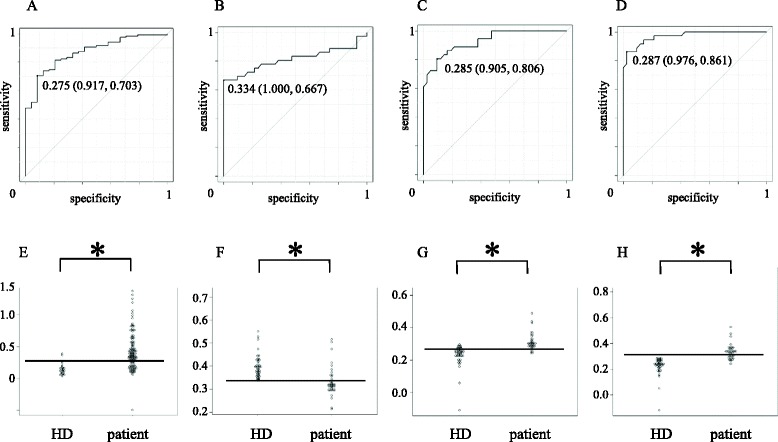
ROC analysis. Receiver operating curve (ROC) analysis for the evaluation of diagnostic value of antibodies against purified RPA2 protein (A), bRPA2-4 (B), bRPA2-90 (C) and bRPA2-132 (D). Areas under the curve (AUC) were 0.867 (95% CI: 0.798-0.936), 0.812 (95% CI: 0.702-0.813), 0.928 (95% CI: 0.874-0.982) and 0.971 (95% CI: 0.940-1.00), for each antigen. Number in the curves indicate cut-off value of RPA2 Ab level and those in the parentheses indicates specificity (left) and sensitivity (right). Dot plot of antibody levels in patients and healthy donors against purified RPA2 protein (E), bRPA2-4 (F), bRPA2-90 (G) and bRPA2-132 (H). The horizontal bar indicates the cut-off value for each antibody level calculated in the ROC analysis. *P < 0.05 by Mann–Whitney U test.
