Figure 3.

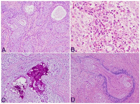
Histological and immunohistochemical features of the tumor. (A) Tumor nests consisted of epithelial cells and pseudoglandular structures (hematoxylin and eosin (H&E) staining; ×40). (B) Marked eosinophilic infiltration in the tumor stroma (H&E; ×200). (C) The tubular structure contained periodic acid–Schiff-positive mucus in the lumen and goblet cells (×100). (D) Venous invasion can also be observed (Victoria blue–H&E; ×100).
