Fig 4.

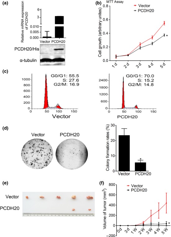
The effect of PCDH20 on HCC cell growth in vitro and in vivo. (a) After selected by G418 for three weeks, the expression level of PCDH20 in stably transfected cell clones was measured by quantitative RT-PCR and Western blot assay, respectively. (b) The growth rate of SNU-449 cells with stable expression of PCDH20 and vector control was measured using MTT assay according to the number of days (d) in culture. Values are presented as the mean ± SD of three independent experiments. *P < 0.05. (c) Cell cycle of SNU-449 cells with ectopic PCDH20 expression was analysed by flow cytometry. (d) Colony formation was measured by soft agar assay. Representative images of colony formed in soft agar and the histograms of relative percentage of colony numbers of the indicated clones were shown. (e, f) Effect of PCDH20 on tumourigenicity in nude mice xenograft model. Tumour volume was measured and plotted as mean ± SD. The curve of tumour growth was shown (n = 5/group).
